Back Bones Diagram / Muscles Of The Lumbar Spine Of The Trunk - Exercises can strengthen the core muscles that support the spine and.. In this image, you will find 1st cervical vertebrae, atlus, cervical plexus, 7th cervical vertebrae, 1st thoracic vertebrae, brachial plexus, spinal dura mater, filaments of spinal nerve roots, 12th thoracic vertebra, 1st lumber vertebra, iliohypogastric nerve, ilioinguinal nerve, lumbar. This spinal column provides the main support for your body, allowing you to stand upright, bend, and twist, while protecting the spinal cord from injury. 5.0 out of 5 stars: The knee joint is the largest joint in the body and is primarily a hinge joint, although some sliding and rotation occur. Arms and hands bones names.
Add to cart add to cart add to cart add to cart customer rating: The red lines point individual bones and the names are writen in singular, the blue lines conect to group of bones and are in plural form. The foot bones shown in this diagram are the talus, navicular, cuneiform, cuboid, metatarsals and calcaneus. It is designed to be incredibly strong, protecting the highly sensitive nerve roots, yet highly flexible, providing for mobility on many different planes. A tough, springy disc of cartilage sits between the vertebrae of your spine.
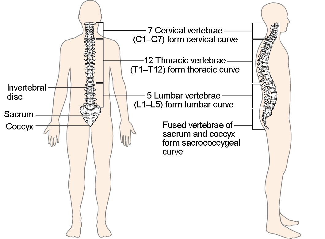
The Vertebral Column Anatomy And Physiology I from s3-us-west-2.amazonaws.com Spine diagram studying a spine diagram is one way to better understand many of the individual components of the back bone and how they might relate to a symptomatic back, neck or sciatica pain condition. Related posts of human back bone chart cross section of human bone diagram. This article looks at the anatomy of the back, including bones, muscles, and nerves. Diagram of a human female skeleton, back view. The bones of the chest and upper back combine to form the strong, protective rib cage around the vital thoracic organs such as the heart and lungs. The vertebrae, which stack like spools of thread, support the back and protect the spinal cord. It also covers some common conditions and injuries that can affect the back. The spine anatomy is a complex structure.
Bones of the pelvis and lower back.
This article looks at the anatomy of the back, including bones, muscles, and nerves. Human body poster 24 x 36in. But, they are common in the back and can cause pain. The bones of the chest and upper back combine to form the strong, protective rib cage around the vital thoracic organs such as the heart and lungs. At the back of each bone in the spine (vertebra) are bony points called processes, which muscles attach to. Bone structure birds 12 photos of the bone structure birds bone structure birds, bone structure in. Diagramme schnell und einfach erstellen. The bones of the pelvis and lower back work together to support the body's weight, anchor the abdominal and hip muscles, and protect the delicate vital organs of the vertebral and abdominopelvic cavities. This spinal column provides the main support for your body, allowing you to stand upright, bend, and twist, while protecting the spinal cord from injury. This item human back bones diagram poster 28 inch x 24 inch / 16 inch x 13 inch. Its appearance is different from the other spinal vertebrae. The lumbar spine connects to the thoracic spine above and the hips below. This vertebra supports the skull.
Add to cart add to cart add to cart add to cart customer rating: Seven cervical vertebrae in the neck, twelve thoracic vertebrae in the torso and five lumbar vertebrae in the lower back. The disks that cushion vertebrae may compress with age or injury, leading to a herniated disk. Atlas (c1) the atlas is the first cervical vertebra and therefore abbreviated c1. Can you feel the bumps of your vertebrae along your back?
Back And Spine Complete Pain Care Helping You Return To You from www.completepaincare.com Each typical vertebra consists of a body, an arch and three processes that stem from. Add to cart add to cart add to cart add to cart customer rating: In this image, you will find 1st cervical vertebrae, atlus, cervical plexus, 7th cervical vertebrae, 1st thoracic vertebrae, brachial plexus, spinal dura mater, filaments of spinal nerve roots, 12th thoracic vertebra, 1st lumber vertebra, iliohypogastric nerve, ilioinguinal nerve, lumbar. Spinal anatomy and back pain. Lateral labeled diagram of the human vertebral spinal column showing vertebrae numbering order and the 5 different regions of the spine. The disks that cushion vertebrae may compress with age or injury, leading to a herniated disk. The atlas is the topmost vertebra, and along with c2, forms the joint connecting the skull and spine. Spinal vertebrae bone spine vertebra toracica spinal cord spine structure back diagram spine sections spinal cord vertebrae spinal structure health diagram.
Arms and hands bones names 12 photos of the arms and hands bones.
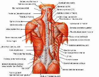
Anatomy of the spine and back spine muscles diagram. A tough, springy disc of cartilage sits between the vertebrae of your spine. Spinal anatomy is a remarkable combination of strong bones, flexible ligaments and tendons, large muscles and highly sensitive nerves. Cross section of human bone diagram 12 photos of the cross section of human bone diagram cross section diagram of human bone, bone, cross section diagram of human bone. At the back of each bone in the spine (vertebra) are bony points called processes, which muscles attach to. The spine diagram the spine diagram shown below, consists of many bones or vertebrae,soft discs,the spinal cord, and spinal nerves. But, they are common in the back and can cause pain. Your lower back contains 5 vertebral bones stacked above each other with intervertebral discs in between. These bones are connected at the back with specialized joints. Atlas (c1) the atlas is the first cervical vertebra and therefore abbreviated c1. The vertebral column is a series of approximately 33 bones called vertebrae, which are separated by intervertebral discs. The bones of the pelvis and lower back work together to support the body's weight, anchor the abdominal and hip muscles, and protect the delicate vital organs of the vertebral and abdominopelvic cavities. Related posts of human back bone chart cross section of human bone diagram.
(temporal bone) shoulder blade (scapula) lower back vertebrae (5) (lumbar vertebrae) back of skull (occipital bone) fused vertebrae (5) (sacrum) hand bones (metacarpals) finger bones Bones, discs, and joints in your lower back. Muscle or tendon injuries can occur anywhere in the body. At the back of each bone in the spine (vertebra) are bony points called processes, which muscles attach to. The knee joint is the largest joint in the body and is primarily a hinge joint, although some sliding and rotation occur.
Spine Basics Orthoinfo Aaos from orthoinfo.aaos.org (temporal bone) shoulder blade (scapula) lower back vertebrae (5) (lumbar vertebrae) back of skull (occipital bone) fused vertebrae (5) (sacrum) hand bones (metacarpals) finger bones Its appearance is different from the other spinal vertebrae. It also covers some common conditions and injuries that can affect the back. Lateral labeled diagram of the human vertebral spinal column showing vertebrae numbering order and the 5 different regions of the spine. The spine supports your body and helps you walk, twist and move. Arms and hands bones names 12 photos of the arms and hands bones. It is designed to be incredibly strong, protecting the highly sensitive nerve roots, yet highly flexible, providing for mobility on many different planes. Female anatomy human body classroom educational chart cool wall decor art print poster 24x36.
The atlas is a ring of bone made up of two lateral masses joined at.
The bones of the chest and upper back combine to form the strong, protective rib cage around the vital thoracic organs such as the heart and lungs. Arms and hands bones names. Atlas (c1) the atlas is the first cervical vertebra and therefore abbreviated c1. Seven cervical vertebrae in the neck, twelve thoracic vertebrae in the torso and five lumbar vertebrae in the lower back. The lumbar spine is the lower back that begins below the last thoracic vertebra (t12) and ends at the top of the sacral spine, or sacrum (s1). The occiput (co), also known as the occipital bone, is a flat bone that forms the back of the head. Arms and hands bones names 12 photos of the arms and hands bones. The spine diagram the spine diagram shown below, consists of many bones or vertebrae,soft discs,the spinal cord, and spinal nerves. The lumbar spine connects to the thoracic spine above and the hips below. Bone diagram forehead (frontal bone) nose bones (nasals) cheek bone (zygoma) upper jaw (maxilla) lower jaw (mandible) breast bone (sternum). Each lumbar spinal level is numbered from top to bottom—l1 through l5, or l6. Bone structure birds 12 photos of the bone structure birds bone structure birds, bone structure in. Female anatomy human body classroom educational chart cool wall decor art print poster 24x36.


0 Komentar